Annals of Medical & Surgical Case Reports
Case Report
Third Degree Atrioventricular Block Following Blunt Chest Injury
Alblooshi A*, Alharmoodi F, Alkaram S and Bekdache O
Department of Surgery and Trauma, Tawam Hospital-in affiliation with John Hopkins, Abu Dhabi, United Arab Emirates
*Corresponding author: Alya Alblooshi, Department of Surgery and Trauma, Tawam Hospital-in affiliation with John Hopkins, Abu Dhabi, United Arab Emirates, Tel: +97150815077; Email: atalblooshi2@gmail.com
Citation: Alblooshi A, Alharmoodi F, Alkaram S, Bekdache O (2019) Third Degree Atrioventricular Block Following Blunt Chest Injury. Ann Med & Surg Case Rep: AMSCR-1000018.
Received date: 22 August, 2019; Accepted date: 04 September, 2019; Published date: 14 September, 2019
Abstract
Blunt chest trauma following motor vehicle collision is a common presentation to the emergency department, however, secondary cardiac complications are considered rare. We are reporting a case of 16-year-old male with third degree atrioventricular block following motor vehicle collision.
Keywords: Atrioventricular block; Blunt Chest Injury; Motor Vehicle Collision
Introduction
Blunt chest injury (BCI) can result from direct trauma to the chest secondary to motor vehicle collisions (MVC); fall down, sport accidents or an animal kick. Resulting cardiac complications could vary from being completely asymptomatic to the development of fatal dysrhythmias and death. The incidence of cardiac complications following trauma was reported to be less than 10% [1], and it is even less frequent to cause complete atrioventricular block (AVB). We are reporting a case of 16-year-old male with no previous cardiac disease, who developed a complete AVB after sustaining a BCI during an MVC [2].
Case Report
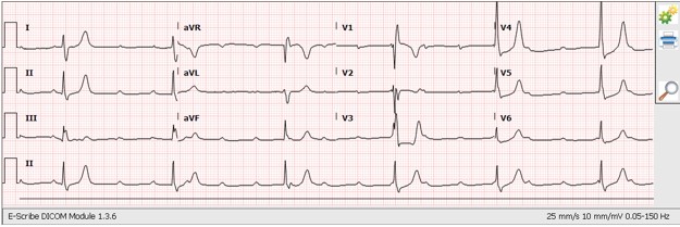
A previously healthy 16-year-old male presented to the emergency room (ER) after being involved in MVC. The patient was an unrestrained front seat passenger. The car rolled over to the driver’s side. He arrived to the ER alert and oriented with a Glasgow Coma Scale of 15, complaining of headache and chest pain. Vital signs showed sinus bradycardia (Figure 1). Initial heart rate readings were ranging between 40s-50s beats per minute (bpm), with normal blood pressure (121/74 mmHg) and respiratory rate (18 breaths/mn) (Figure 1).
Electrocardiogram (ECG) on arrival to our ER showed Sinus tachycardia, third degree AV block, with probably a junctional escape rhythm with incomplete right bundle branch block (RBBB).
Computerized Tomography (CT) of the head, chest, abdomen and pelvis showed a small right sided apical pneumothorax, end plate fracture of thoracic vertebra 11, scalp hematoma and possible Grade1 splenic injury. There were no signs of pericardial effusion, cardiac injury, or rib fractures.
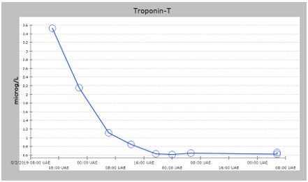
Initial abnormal laboratory findings showed troponin level of 3.5 microgram/L (normal high <=0.014), haemoglobin of 155 gm/L, White Blood Cell count (WBC) of 26.7 ×10^9/L and Lactate of 3.0 mmol/L.
Echocardiogram (Echo) revealed normal left ventricle (LV) size, good LV systolic function, mildly dilated Right ventricle (RV) with mild hypokinesia. Mild to moderate Tricuspid Regurgitation (TR), significant pulmonary hypertension (SPAP ? 60 mmHg) were also noted. No clots or pericardial effusion identified. Cardiology recommended CT chest with angiogram to rule out de novo thrombotic occlusion in the pulmonary arteries. It turned out to be negative.
Patient was admitted to the intensive care unit (ICU) for observation with serial ECGs, Echos and troponin levels. Levels were trending down during the observation period (Figure 2).
Patient remained in ICU in a stable condition, while still having bradycardia with heart rate of (30s-50s bpm) and normal blood pressure. Follow up ECG finding remained the same with picture of complete hearth block (Figure 1).
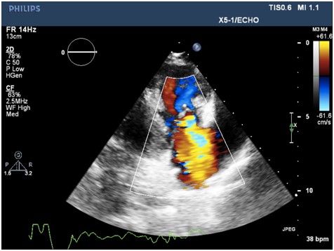
Repeated Echo scan showed severe TR, multiple jets with possibility of secondary leaflet perforation, a congenital cleft, or a prolapse due to the trauma itself. Moderate pulmonary hypertension was seen unchanged. Mildly dilated RA and RV with Normal RV function were also noted (Figure 3).
Decision was made to transfer the patient to a higher facility with available cardiac surgery service after temporary pacing to ensure the safety of transfer. Transcutaneous temporary pacemaker was inserted uneventfully using a right Jugular vein access into the right ventricle.
The Patient was transferred in good general condition. Repeated Echo again showed severe TR, speculated to be caused by a prolapse of all the tricuspid valve leaflets, with flail seen at the septal leaflet, caused by severe and high gradient flow originating from a ventricular septal rupture (VSR) noted at the membranous inter-ventricular septum, just below the aortic valve annulus. CT Cardiac Angiogram was done to evaluate for congenital cardiac anomalies, it showed a musclomembranous VSD. The demonstrated VSD was likely congenital and incidentally diagnosed during this presentation given the patient’s hemodynamic stability.
The patient was discharged in a good general condition. Follow-up was done after one week in a specialized adult congenital heart disease clinic. The patient was noted to be asymptomatic with good general condition. Repeated Echo was similar to the last scan. Repeated ECG was showing sinus rhythm and right bundle branch block (RBBB).
The patient will continue to follow in the adult congenital heart disease clinic regularly with clear instructions to maintain an excellent oral hygiene with regular visits to the dentist. He was encouraged to maintain a healthy active lifestyle with no exercise limitations, as per the recommendations of the American Heart Association [3].
Discussion
Cardiac involvement during BCI needs to be suspected in the setting of severe anterior chest impact, symptomatic patients with dysarrhythmias, and abnormal cardiac enzymes. ECG abnormalities need to be addressed by a specialized physician to decide for further diagnostic modalities, such as transthoracic Echo, trans-oesophageal Echo (TOE) or conventional angiography. None of the previously mentioned modalities was found to be accurate enough to diagnose cardiac contusion or to predict all complications. Latest studies have shown that troponin I and troponin T can help in the stratification of patients at risk for complications [4-7].
Risk stratification of patients with blunt cardiac injury can be done according to hemodynamic stability and initial markers, which could help direct the disposition of the patient and the need for further workup.
In cases of detected cardiac injuries, patients should be closely monitored in the early stages to treat any adverse events, patients usually need to be monitored closely in the first 24-48 hours, as most complications tend to occur during this period, and any further management needs to be performed in adequate settings equipped with full spectrum of cardiac multidisciplinary services.

Figure 1: Vital signs showed sinus bradycardia.

Figure 2: Levels were trending down during the observation period.

Figure 3: Mildly dilated RA and RV with Normal RV function were also noted.
Citation: Alblooshi A, Alharmoodi F, Alkaram S, Bekdache O (2019) Third Degree Atrioventricular Block Following Blunt Chest Injury. Ann Med & Surg Case Rep: AMSCR-1000018.