
Journal of Surgery and Insights
Free Tubercular Illeal Perforation with Ascaris Coinfection: A Rare Case Report
Anant Pore1, Ankush Sarwal1*, Pulkit Maru2 and Neetika Sarwal3
1Department of Surgical oncology, Tata Medical Centre, Kolkata, India
2Department of Radiodiagnosis, Tata Medical Centre, Kolkata, India
3Department of Anaesthesiology, MMIMR, Mullana, India
*Corresponding author: Dr. Ankush Sarwal, Department of Surgical oncology, Tata Medical Centre, 14 MAR (E-W), New Town, Rajarhat, Kolkata, India. Tel: +919812636877, Email: drankushsarwal@yahoo.in
Citation: Pore A, Sarwal A, Maru P, Sarwal N (2019) Free Tubercular Illeal Perforation with Ascaris Coinfection: A Rare Case Report. J Surg Insights: JSI-100002.
Received Date: 04 May, 2019; Accepted Date: 09 May, 2019; Published Date: 16 May, 2019
Abstract
Acute abdomen encompasses around 5-10% of emergency department admissions. Although peptic ulcer perforations account for the majority of cases of peritonitis, small bowel perforations are also commonly encountered. Tuberculosis remains an uncommon cause of perforation of the small bowel. The surgical procedure for management depends on site and extent of disease, status of the remaining gut, general condition of the patient, surgeon’s expertise and individual preference. We report a case of a free tubercular perforation of the ileum with secondary infection Ascaris Lumbricoides that presented with peritonitis and was managed at our hospital.
Keywords: Acute abdomen; Ascaris Lumbricoides; Tubercular peritonitis
Introduction
Perforation of a hollow viscus leading to peritonitis is a common surgical emergency. Although peptic ulcer perforations account for the majority of cases of peritonitis, small bowel perforations are also commonly encountered. Usually, these small bowel perforations are secondary to enteric fever or trauma, but at times, non- specific ileal perforations are also seen. Tuberculosis remains an uncommon cause of perforation of the small bowel [1,2]. We report a case of a free tubercular perforation of the ileum with secondary infection Ascaris Lumbricoides that presented with peritonitis and was managed at our hospital.
Case Summary
The patient was an18-years old female who presented with complaint of generalized pain in abdomen with multiple episodes of bilious vomiting, fever for 1 week to emergency department. On examination she was ill and septic, the abdomen was distended and muscular resistance could be felt. Ultrasonography (USG) of Abdomen was done which showed Moderate amount of free fluid. Computed Tomography (CT) Abdomen was done which showed Pneumoperitoneum with distal ileal obstruction. CT showed mottled small bowel loop with segmental stricture, with leak seen in distal illeum and dilatation with moderate ascites and peritoneal thickening and enhancement. TB gold test was sent which came out to be positive. Informed Consent for emergent laprotomy was taken and patient was shifted to operating room. Emergent laparotomy was performed in which purulent ascites was found. Intestinal perforation was seen in distal ileum around 2 feet from ileo-cecal junction along with dense adhesions in intestines. Resection of ileum along with end to end anastomosis was done. Post operatively patient was shifted to surgical ICU, started on IV fluids and Anti-tubercular medicines and Total parentral nutrition (TPN). Patient had purulent discharge from the operative site on post-operative day (POD) 3 suggesting burst abdomen. On inspection the operative site on POD 4 fecal matter was seen coming from the abdominal wound. As she was hemodynamically stable and fecal fistula output was less than 100 ml, she was managed conservatively with IV Antibiotics, Anti Koch’s Treatment and TPN (Figure 1). On POD 9, a large worm suggesting to be Ascaris Lumbricoides was recovered from abdominal wound during dressing (Figure 2). Hence patient treatment was started on antihelmenthics which was Albendazole 400 mg twice daily for 1 week. Patient was doing fine and was started on oral soft diet. But, on POD 15patient`sabdominal distention increased along with deterioration in hemodynamics. An urgent CT Abdomen and Pelvis was ordered which was showed centrally clumped and adherent small bowel loops with ill-defined diffuse wall thickening involving jejunum and ileum up to visualized distal ileum, abdominal wall dehiscence involving supraumbilical and infraumbilical region with low density collection within subcutaneous tissue and enterocutaneous fistula communicating between distal ileum and anterior abdominal wall with active leak of orally administered contrast within subcutaneous space and no further passage of oral contrast. Based on these CT findings Patient was then shifted for emergent Laparotomy. Intraoperatively patient was found to have multiple perforations in distal ileum, so resection of 30 cms of ileum with side to side ileocolic anastomosis was done. Post operatively patient was shifted to surgical ICU, given IV fluids, IV antibiotics and parentral nutrition. Patient recovered well and was started on Liquid diet on POD 4. She was shifted to ward on POD 5. Her Anti-tubercular therapy (ATT) and Anti helminthic treatment was shifted to oral medications. She was discharged on POD12 and has now completed her 12 months of ATT.
The specimen was sent for histopathological examination and the final histopathological examination showed granulomatous inflammation with caseating granulomas along with organized serositis suggestive of tuberculosis.
Discussion
Primary intestinal tuberculosis (without pulmonary involvement) is one of the commonest forms of extrapulmonary tuberculosis. The infection is usually caused by ingestion of unpasteurized or contaminated milk that leads to a primary infection of the intestine in the absence of pulmonary disease [1]. Intestinal tuberculosis commonly affects the ileocaecal region because of the following reasons: 1) the terminal ileum is an area of physiological stasis; 2) it has abundant lymphoid tissue; and 3) it has a high absorptive capacity. Thus, after the initial infection occurs in the Peyer's patches, mucosal edema and sloughing occur, leading to the formation of typical tubercular ulcers that lie transversely to the long axis of the ileum. The disease may spread further by dissemination through the lymphatics and by caseation, may heal by fibrosis, or may even remain confined to the area if the host's defence mechanisms are adequate [3,4].
There are three main pathological forms of tubercular enteritis: 1) ulcerative, 2) hypertrophic, and 3) ulcerohypertrophic. The ulcerative form of the disease is more common than the others, but these ulcers rarely perforate. Fibrosis and the formation of adhesions to adjacent intra-abdominal organs account for the low incidence of perforation seen in tuberculosis. However, if perforation occurs, the patient presents with the signs and symptoms of peritonitis. Although it is well documented that the incidence of perforation in patients with intestinal tuberculosis varies from 1% to 11%, the majority of these perforations (70%-80%) are not truly perforations of such tubercular ulcers, but are ‘blow outs’ of the small bowel secondary to distension due to distal obstruction (strictures or adhesions). As such, true or ‘free’ perforations are rare, and only a few cases have been reported to the present in the world literature, with an overall mortality rate of nearly 70%. Recently, vasculitis of the mesenteric vasculature due to tuberculosis has been implicated as a contributory factor, but the exact mechanism by which some patients develop perforation and others is not established [3-5].
The treatment of tubercular peritonitis is similar to that for peritonitis due to other causes like resuscitation, nasogastric aspiration, intravenous fluids, antibiotics, and surgery once the patient is stabilized. Tubercular perforation is rarely diagnosed pre-operatively as the signs and symptoms are similar to those of peritonitis and there are no pathognomic features either on investigation or on clinical examination. Even in patients who are known to be sufferers of the disease, the diagnosis of perforated tubercular ulcers cannot be made with certainty. The treatment of the perforation depends upon the condition of the patient and the bowel. Primary closure of the perforation can be considered safe if the patient has presented early and the bowel is healthy, otherwise, exteriorization of the affected bowel as a loop ileostomy is a safer option. If there is a long segment of bowel that is diseased, or there are multiple perforations, resection with either primary anastomosis or exteriorization may be considered [3-5].
Intestinal parasitic infections are among the major public health problems in developing countries. Hence, it is significant to explore coinfection with intestinal parasites and tuberculosis because coinfection increases the complexity of control and prevention of tuberculosis and parasitic diseases. Once biopsy confirms the diagnosis of tuberculosis of the bowel, anti-tubercular therapy is mandatory along with anti-helminthics in the present case.
Conclusion
Abdominal Tuberculosis is very difficult to diagnose and diagnosis is often delayed till an acute abdomen is presented with. The most common pathology is intestinal stricture with or without perforation. Almost all patients need surgical intervention. Prompt surgical exploration, vigilant postoperative care and administration of AKT helped to treat the patients successfully with their complete cure and rehabilitation. In case of co-infection with parasite, anti helmenthics also needs to be given. The choice of surgical procedure depends on site and extent of disease, status of the remaining gut, general condition of the patient, surgeon’s expertise and individual preference.
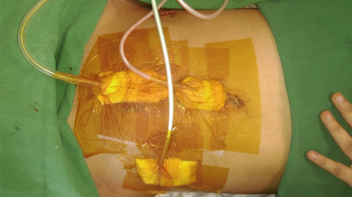
Figure 1: Conservative management of fecal fistula.

Figure 2: Ascaris Lumbricoides from operative wound site.
Citation: Pore A, Sarwal A, Maru P, Sarwal N (2019) Free Tubercular Illeal Perforation with Ascaris Coinfection: A Rare Case Report. J Surg Insights: JSI-100002.
