
Annals of Medical & Surgical Case Reports
(ISSN: 2652-4414)
Image Article
Esophageal Candidiasis
Meng-You Zeng1,2, Bo-Juan Lang3,4, Wei Liu*1,2
1Institute of Digestive Disease, China Three Gorges University, Yichang, China 2Department of Gastroenterology, Yichang Central People’s Hospital, Yichang, China 3Institute of Pathology, China Three Gorges University, Yichang, China
4Department of Pathology, Yichang Central People’s Hospital, Yichang, China
*Corresponding Author: Wei Liu, Institute of Digestive Disease, China Three Gorges University, 8 Daxue Road, Yichang 443000, China
Citation: Zeng YM, Lang BJ, Liu W (2020) Esophageal Candidiasis. Ann Med & Surg Case Rep: AMSCR-100087
Received date: 12 December 2020; Accepted date: 23 December 2020; Published date: 29 December 2020
A 57-year-old man presented to the outpatient clinic with a 2-week history of odynophagia (pain on swallowing). His past medical history was a sufferer of asthma of 50 years’ duration with twice-daily budesonide inhalation suspension for the treatment of persistent asthma. Physical examination was unremarkable.Gastroscopy revealed white, linear, plaquelike, mucosal lesions on the esophagus (Panel A). No oropharyngeal and gastroduodenal lesions were found. Biopsy of mucosa confirmed Candida blastospores and pseudohyphae (Panel B). Direct fungal cultures and detection were also positive for Candida albicans and a serologic assay for human immunodeficiency virus was negative. The diagnosis of esophageal candidiasisdepended on the typically characteristic endoscopic manifestations and was confirmed by a culture of esophageal brushing samples which was positive for Candida albicans [1]. Esophageal candidiasis is considered to be a common opportunistic infection in immunocompromised hosts [2]. Long-term treatment with glucocorticoidsis a risk factor in immunocompetent patients [3]. Oral fluconazole was administered for 3 weeks, and his pain on swallowing relieved within 1 month after treatment was initiated.No additional endoscopy was performed after completion of the procedure. At a 1-year follow-up visit, the patient reported no further symptoms of odynophagia.
Acknowledgements
Funding: This work was supported by National Natural Science Foundation of China (31600134).
Conflicts of Interest: The authors have no conflicts of interest to declare.
Ethical Statement: The authors are accountable for all aspects of the work in ensuring that questions related to the accuracy or integrity of any part of the work are appropriately investigated and resolved. Written informed consent was obtained from the patient for publication of this “Images in Clinical Medicine”.
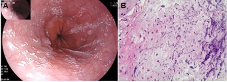
Figure 1: Esophageal candidiasis. (A) Endoscopic appearance of white mucosal plaques in lower esophagus; (B) Candida blastospores and pseudohyphae detected by biopsy of mucosa
Citation: Zeng YM, Lang BJ, Liu W (2020) Esophageal Candidiasis. Ann Med & Surg Case Rep: AMSCR-100087
