
Annals of Medical & Surgical Case Reports
(ISSN: 2652-4414)
Image Article
A Patient with Gastric Schwannoma Mimicking Gastrointestinal Stromal Tumor
Meng-You Zeng1, 2, Lu-Gao Tian1, 2, Bo-Juan Lang3, 4, Yuan Yuan3, 4, Wei Liu*1, 2
1Institute of Digestive Disease, China Three Gorges University, Yichang, China
2Department of Gastroenterology, Yichang Central People’s Hospital, Yichang, China
3Institute of Pathology, China Three Gorges University, Yichang, China
4Department of Pathology, Yichang Central People’s Hospital, Yichang, China
*Corresponding Author: Wei Liu, Institute of Digestive Disease, China Three Gorges University, 8 Daxue Road, Yichang 443000, China
Citation: Zeng MY, Tian LG, Lang BJ, Yuan Y, Liu W (2020) A Patient with Gastric Schwannoma Mimicking Gastrointestinal Stromal Tumor. Ann Med & Surg Case Rep: AMSCR-100085
Received date: 12 December 2020; Accepted date: 23 December 2020; Published date: 29 December 2020
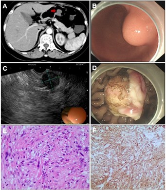
A 48-year-old woman underwent computed tomography (CT) due to epigastric pain. The CT scan revealed a 21 × 17 mm gastric wall mass protruding into the stomach cavity (Figure A, arrow). Gastroscopy confirmed a 2-cm submucosal mass in the greater curvature (Figure B). and mucosal biopsy was unremarkable. Endoscopic ultrasound found an endophytic- exophytic hypoechoic mass with heterogeneous texture arising from the muscularis propria (Figure C). The nodule was removed using a snare afterendoscopic submucosal dissection (Figure D). The histology confirmed moderately cellular spindle-cell tumor (Figure E) and immunostaining of the tumor cells was positive for S100 (Figure F) and negative for DOG1, CKIT, SMA, and desmin, all consistent with a schwannoma arising from the muscularis mucosa instead of gastrointestinal stromal tumor. As very rare neoplasms of the stomach, schwannomas are mostly asymptomatic and discovered incidentally with exceedingly rare malignant transformation [1, 2]. The overall prognosis of these lesions is unknown. The treatment usually includesendoscopic or surgical resection with the diagnosis mostly made postoperatively by immunohistochemistry [1]. Therefore, we need to consider schwannomas in the differential diagnosis of submucosal gastric tumors of the muscularis propria layer. Surgical excision with negative margins is the recommended treatment of choice because incomplete excision may be relevant with local recurrence or distant metastasis. Endoscopic mucosal resection with intermittent endoscopic surveillance and long-term clinical follow-up evaluation is suggested for small gastric schwannomas because of the rare risk of malignant transformation.We are planning to repeat endoscopic surveillance in 1 year for our patient.
Acknowledgements
Funding: This work was supported by National Natural Science Foundation of China (31600134).
Conflicts of Interest: The authors have no conflicts of interest to declare.
Ethical Statement: The authors are accountable for all aspects of the work in ensuring that questions related to the accuracy or integrity of any part of the work are appropriately investigated and resolved. Written informed consent was obtained from the patient for publication of this “Images in Clinical Medicine”.
Citation: Zeng MY, Tian LG, Lang BJ, Yuan Y, Liu W (2020) A Patient with Gastric Schwannoma Mimicking Gastrointestinal Stromal Tumor. Ann Med & Surg Case Rep: AMSCR-100085
