
Journal of Obstetrics and Gynecological Problems
Case Report
Prenatal Diagnosis of Fetal Goiter in a Euthyroid Mother Under Chemotherapy for Breast Cancer: Is There a Link Between the Two Conditions?
Alpha boubacar C*, Aboubecrine B, Zohra fdili AF, Sofia J, Abraham Alexis S, Hikmat C and Abdelillah MM
Department of Gynecology-Obstetrics II, Hassan II Teaching Hospital, Sidi Mohamed Ben Abdellah University, Fez Morocco
*Corresponding author: Conte Alpha boubacar, Department of Gynecology-Obstetrics II, Hassan II Teaching Hospital, Sidi Mohamed Ben Abdellah University, Fez , Morocco
Citation: Alpha boubacar C, Aboubecrine B, Zohra fdili AF, Sofia J, Abraham Alexis S, et al. (2020) Prenatal Diagnosis of Fetal Goiter in a Euthyroid Mother Under Chemotherapy for Breast Cancer: Is There a Link Between the Two Conditions?. J Obstet Gynecol Probl: JOGP 100016
Received date: 05 September, 2020; Accepted date: 10 September, 2020: Published date: 15 September, 2020
Abstract
Congenital goiter is an enlarged thyroid gland in newborns, and may be associated with hyperthyroidism, hypothyroidism, or even euthyroidism. The incidence of congenital hypothyroidism is estimated to one in every 40,000 live births and is one of the most common treatable causes of mental retardation. The diagnosis is not evident when the mother is known free of any thyroid dysfunction. We described in this manuscript a prenatal diagnosis of fetal goiter in a thyroid mother under chemotherapy for breast cancer. We also made a review of the literature to find if the link between the two conditions has ever been reported or proved.
Keywords: Chemotherapy; Euthyroid mother; Fetal goiter
Introduction
Congenital goiter is an enlarged thyroid gland in newborns, and may be associated with hyperthyroidism, hypothyroidism, or even euthyroidism [1]. Fetal thyroid disorders are often unrecognized when there is no maternal history of thyroid disease [2]. The incidence of congenital hypothyroidism is estimated to one in every 40,000 live births and is one of the most common treatable causes of mental retardation [3]. Fetal hypothyroidism causing fetal goiter can lead to polyhydramnios due to esophageal compression, hyperextension of the neck leading to dystocia during labor and may cause compression of the airway, thereby causing asphyxia [4] Sonography is very useful at detecting fetal neck masses, and the use of 3D technology provides an in-depth evaluation of the mass. Fetal goiters can be easily detected in the sagittal profile view of the fetus. When a sonographer detects a fetal goiter, it is important to document the size and vascularity of the goiter [5,6]. There is possibility of in utero treatment.
Case report
35-year-old patient with no notable pathological history and no family history of goiter G6P5 using estroprogestative contraception for more than 10 years who consulted for the management of a nodule in the left breast diagnosed as carcinoma treated by chemotherapy based on FEC (Fluorouracil/Epirubicin/Cyclophosphamide) and Docetaxel. She reported the notion of 4-months amenorrhea in who an obstetric ultrasound discovered an active mono-fetal pregnancy whose gestational age was estimated at 16 weeks + 5 days without any abnormal morphology finding. The pregnancy was monitored by regular checkup. The obstetric ultrasound done at 28 weeks found a neck anterior mass. The mass was homogenous and echoic with vascularization and without other visible abnormalities.
The maternal hormonal balance was also normal. We therefore concluded that there was a fetal goiter in a patient without a thyroid abnormality carrying breast cancer under chemotherapy. We continued to monitor the fetus by doing regular ultrasonography researching other signs like hydramnios until the 37th week. The patient delivered naturally and the newborn died during the first hour of life.
Discussion
Fetal goiter occurring to a euthyroid mother is a rare event. The diagnosis is often unrecognized when there is no maternal history of thyroid disease. Sometimes, a fetal goiter is difficult to detect by ultrasound when the condition is not severe, and must be carefully examined during routine evaluation. When a fetal goiter is detected during antenatal examination, a detailed evaluation including maternal history and maternal physical examination (heart rate, heat intolerance, weight change, exophthalmos, etc.) is necessary [2].
According to [1] fetal goiter is considered a rare occurrence during intrauterine development, with an incidence of 1:40,000 cases. Fetal thyroid hypertrophy leads to three possible associations: hyperthyroidism, hypothyroidism and euthyroidism. Consequently, assessment of thyroid hormone function is of great importance for guiding early treatment. Fetal goiters can arise from maternal grave’s disease, Hashimoto thyroiditis, iodine ingestion, propylthiouracil exposure, and methimazole exposure. They can also be caused from primary fetal hypothyroidism [5,6]. In our present report, there were no maternal thyroid pathological story neither other known etiology nor exposure apart from the fact that the patient was under chemotherapy regiment (FEC and Docetaxel) for the management of her breast cancer. This kind of situation has not been previously observed in our department where a recent study made on pregnancy associated breast cancer did not find any abnormalities in fetus despite different regimens of chemotherapy [7].
Fetal goiters can lead to neck hyperextension, polyhydramnios and compression of the vascular structures and other structures in the neck with motor and mental retardation at a later stage, making the early diagnosis and appropriate management of fetal goiter important. [8, 9]. At delivery, respiratory compromise can also occur as a result of trachea compression by the enlarged thyroid gland [3]. The newborn of our patient died during the first hour of life in a condition we could not described as the patient choosed to deliver elsewhere than our health care center.
Ultrasound score proposed includes fetal heart rate, fetal movements, color doppler for vascularity and bone maturation. Color Doppler can indicate fetal thyroid function with the pattern of vascularity. Peripheral vascularity suggests non?functioning hypertrophied gland whereas central vascularity suggests overactive hyperfunctioning gland [10].
On suspicion of hypothyroidism, the assessment of thyroid function for potential intrauterine treatment is essential to avoid long-term neurological outcomes [11]. To reduce the risk of neonatal death because of tracheal obstruction and mechanical issues at delivery caused by fetal neck hyperextension the treatment of a large fetal goiter should be the priority once the diagnosis is confirmed (Figure 1).
This intrauterine treatment prevents also the possibility of premature rupture of membranes and preterm delivery due to polyhydramnios and dystocia in normal labor due to fetal neck hyperextension. Normal intrauterine growth with normal CNS development can be achieved in euthyroid metabolic state [11]. According to Davidson KM and al, once fetal hypothyroidism is diagnosed, it can be treated with direct administration of thyroid hormones to the fetus, with intra-amniotic administration of synthroid being the least invasive approach to fetal treatment.
Conclusion
Prenatal diagnosis of fetal goiter in a thyroid mother is rare. Despite its rarity the diagnosis is possible with good ultrasound. Some causes have been reported in the literature. Our patient was carrying a breast cancer which was managed by chemotherapy. The link between her chemotherapy regiment and the occurrence of fetal goiter has not been previously described. In this present case, the link has also not been proved.
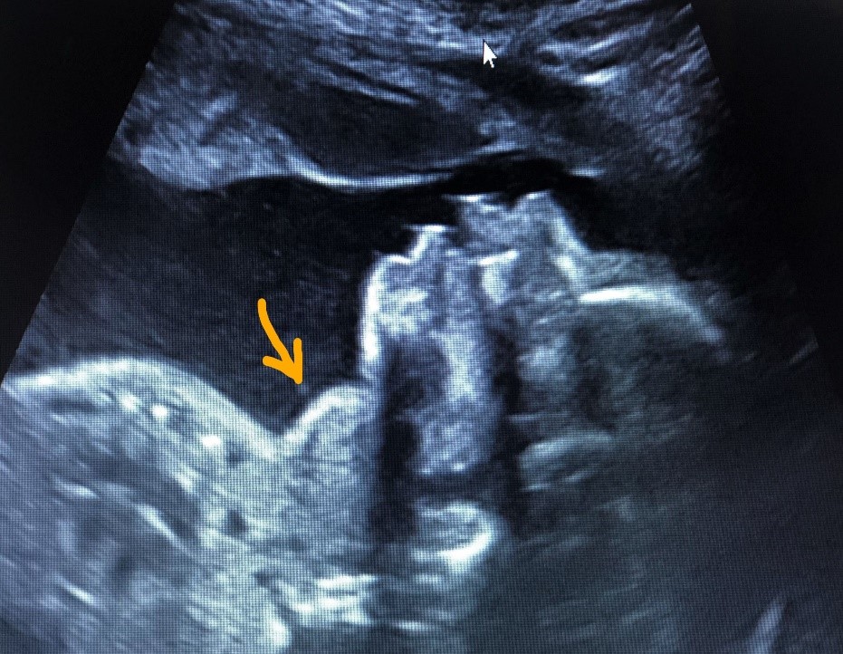
Figure 1: Cervical mass described as fetal goiter.
Citation: Alpha boubacar C, Aboubecrine B, Zohra fdili AF, Sofia J, Abraham Alexis S, et al. (2020) Prenatal Diagnosis of Fetal Goiter in a Euthyroid Mother Under Chemotherapy for Breast Cancer: Is There a Link Between the Two Conditions?. J Obstet Gynecol Probl: JOGP 100016
