
Annals of Medical & Surgical Case Reports
(ISSN: 2652-4414)
Case Report
A Patient with Hobnail Hemangioma-A Case Report
Almadani H and Baeasa IT*
Department of Pediatrics, King Fahad Armed Forces Hospital, Jeddah, Kingdom of Saudi Arabia, Saudi Arabia
*Corresponding author: Dr. Housam Almadani, Department of Pediatrics, King Fahad Armed Forces Hospital, Jeddah, Kingdom of Saudi Arabia, Saudi Arabia.
Citation: Almadani H and Baeasa IT (2020) A Patient with Hobnail Hemangioma-A Case Report. Ann Med & Surg Case Rep: AMSCR- 100070.
Received date: 14 July, 2020; Accepted date: 21 July, 2020; Published date: 27 July, 2020
Targeted Hemosiderotic Hemangioma (THH), also known as Hobnail hemangioma. Is a lesion of probably lymphatic or vessels in origin. With characteristic clinical and histological features. The most clinical feature is small single solitary violaceous papule with targeted-appearing lesion. Histopathologically, there is a biphasic pattern with superficial proliferation of ectatic dermal vascular lumina with intraluminal papillary projections, and pseudoangiosarcomatous pattern in the deep dermis, and endothelial cells are flat or conspicuously epithelioid with solid intraluminal projections giving the hobnail morphology. A simple excision is curative.
Keywords: Hobnail hemangioma; Hemosiderotic hemangio-ma; Superficial hemosiderotic malformation; Targeted
Introduction: Targetoid Hemosiderotic Hemangioma (THH), also known as Hobnail hemangioma, it’s mostly lymphatic origin small solitary benign vascular lesion [1]. That seen in extremities and rarely trunk, of the second decade of life [2]. It was named based on the lesion’s feature histologically [3]. In 1988, Santa Cruz and Aronberg [4] described hobnail hemangioma according the clinical feature and the vascular lesion. Then in 1994, [5] described it and differentiated for the benign vascular lesion regardless the presence of clinical feature. Nevertheless, we have no pediatric cases report it in our area.
Case report
A 1-month old girl full term patient, born in Jeddah she had been well, presented with history of small papule skin lesion in right lower limb since 3rdday of life (Figure 1) that became multiple papulear ulcerated desquamated lesion mainly in right lower limb at 3rd week of life (Figure 2). No history of trauma or known medical condition. The mother sought medical advice and the patient received 1 week of local steroid with no improvement, the lesion increased in size 1 × 2 cm and 2 × 2 cm and became more ulcerative, extensive and with pus and bloody discharge.
Laboratory test including CBC, renal profile, liver profile, coagulation profile, nitrobluetetrazolium test and immunoglobulin level were all within normal ranges. Full septic done and came negative, wound culture showed klebsilla pneumonia in which patient started on antibiotic for total of 14 days. ECHO, ultrasound abdomen and Doppler ultrasound for the lower limb were unremarkable. MRI\MRV\MRA showed mainly artifact which is also unremarkable in our case. Dermatology consulted and biopsy taken from the lesion which showed The papillary dermis ectatic blood vessels lined by bland endothelial cells with hobnail appearance (Figure 3,4).
Discussion
The present case showed small targeted lesion, with central papule, and red halo surrounding. Microscopic examination demonstrates biphasic pattern with dilated vessels lined by hobnail appearance of the endothelial cells with papillary projection and presence of extravasation of the erythrocytes. Therefore, diagnosis of Targetoid Hemosiderotic Hemangioma (THH) were established.
Targetoid Hemosiderotic Hemangioma (THH) presenting classically in stages, early stage of THH presented as targetoid small, single, red lesion with hemorrhagic halo [6]. Later stage only central papule will remain there and the halo may disappear [6]. Most cases the lesion located in the trunk and extremities with no gender predominance. And affected 2nd decade of life [7].
Although Trauma is one of the main cause for Targetoid Hemosiderotic Hemangioma (THH), still the exact pathogenesis is unknown. The clinical presentation of Targetoid Hemosiderotic Hemangioma (THH) varies. This variation with same histologic features explain why THH confused clinically in diagnosis with hemangioma, dermatofibroma, reaction to insect bite and melanoma [5], in which dermocopy good in differentiating between them.
Histologically it varies according to the extent of the biopsied lesion. Papillary dermis and dilated vessels endothelial cells and intraluminal projection with hobnail appearance mostly in initial stages [5], sometimes with extensive red blood cell extravasation, fibrin thrombi and lymphocytic inflammation. With collapsed vascular lumen and stroma hemosiderin deposition in late stages.
Targetoid Hemosiderotic Hemangioma (THH) benign, rare lesion that may mimic malignancy in histological finding. Kaposi’s
sarcoma may have simulated by erythrocyte extravasation and hemosiderin deposition with collagen bundles that dissected by vessels, angiokeratoma simulated by superficial dermis dilated vessels with angiomatous proliferation.
To identify the origin of this hemangioma, it is unclear whether it’s arises from lymphatic vessels endothelial cells or from blood vessels endothelial cell. Immunohistochemically studies will differentiate between them by doing CD31 and CD34 for blood vessels origin and D2 with D40 for lymphatic vessel origin were both negatives in outpatient.
Conclusion
As Targetoid Hemosiderotic Hemangioma (THH) is a benign lesion it’s only removed for cosmetic reasons or correct histological diagnosis of the lesion. Simple excision is therapeutic and diagnostic.

Figure 1,2: History of small papule skin lesion in right lower limb since.
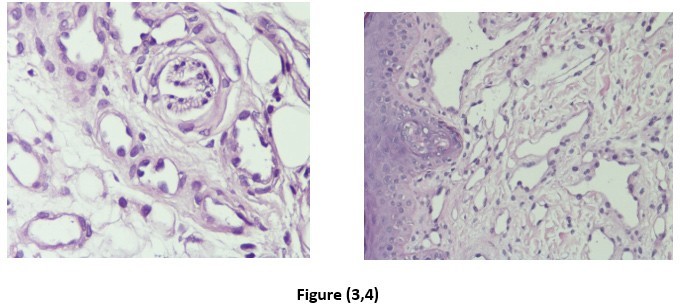
Figure 3,4: Hobnail appearance.
Citation: Almadani H and Baeasa IT (2020) A Patient with Hobnail Hemangioma-A Case Report. Ann Med & Surg Case Rep: AMSCR- 100070.
