
Annals of Medical&Surgical Case Reports
(ISSN 2652-4414)
Research Article
Darier Ferrand Sarcoma: Clinical and Therapeutic Aspects About 8 Cases
FobaL*,Sankale Anne-Aurore,Ndiaye Ai and Ndiaye L
Department of Plastic and Aesthetic Surgery, Aristide Le Dantec University Hospital, Dakar, Senegal
*Corresponding author: Lassana Foba, Department of Plastic and Aesthetic Surgery, Aristide Le Dantec University Hospital, Dakar, Senegal.
Citation: Foba L, Sankale Anne-AuroreNdiaye A, Ndiaye L (2020) Darier Ferrand Sarcoma: Clinical and Therapeutic Aspects About 8 Cases. Ann Med &Surg Case Rep: AMSCR-100058
Received date: 01 May, 2020; Accepted date: 05 May,2020; Published date: 12 May, 2020
Abstract
Background: The dermatofibrosarcoma of Darier and Ferrand is a spindle-shaped cells dermic tumor of intermediate malignancy, characterized by a slow evolution with a major risk of recurrence in case of insufficient resection. With an intermediate malignancy, this tumor is neither strictly malignant (it never metastasizes), nor strictly benign (it is characterized by its strong tendency to recurrence in case of insufficient resection). Not too frequent, our goal is to report our experience in the management of this pathology.
Patient and Methods: It is a retrospective study over a period of 15 years from January 2004 to July 2019 concerning 8 patients cared for in the Plastic Surgery Department of the Aristide Le Dantec Hospital.
Results: Our patients are young with a middle age of 31 years [22 years - 54 years] with as many women as men. Patients consult late on average 5.3 years after the onset of the tumor. Six patients over 7 or 85.7% had a successful first resection. The predominant location of lesions was the shoulder in 4 patients over 7 or 57.1%. The other locations were at the temporal level of the cheek and at the buttock. Histology was obtained on the excision banks, and has noticed healthy margins. The histology of the operative parts confirms the dermatofibrosarcoma of Darier and Ferrand.
Conclusion: The dermatofibrosarcoma of Darier and Ferrand is a malignant tumor of variable location whose management is essentially surgical with wide excision margins to prevent recurrence.
Keywords: Clinical and Therapeutic Aspects; Cutaneous tumor; lymphadenopathy; Patients
Introduction
The dermatofibrosarcoma of Darier and Ferrand is a spindle-shaped cells dermic tumor of intermediate malignancy, characterized by a slow evolution with a major risk of recurrence in case of insufficient resection. [1-8]. It has been described by Darier and Ferrand in 1924 under the name of progressive and recurrent dermatofibroma, called dermatofibrosarcoma protuberans by Anglo-Saxons [7](Figure 1).
With an intermediate malignancy, this tumor is neither strictly malignant (it never metastasizes), nor strictly benign (it is characterized by its strong tendency to recurrence in case of insufficient resection).Not too frequent, our goal is to report our experience in the management of this pathology(Figure 2).
Materials and Methods
It is a retrospective study over a period of 15 years from January 2004 to July 2019 concerning 8 patients cared for in the Plastic Surgery Department of the Aristide Le DantecHospital(Figure 3).
Inclusion criteria: Patients that went under surgery in the cosmetic and reconstructive plastic surgery department, carriers of a dermatofibrosarcoma with histological confirmation.
Exclusion criteria: Patients that have not undergone surgery in the department, patients having other cutaneous tumor different from the Darier-Ferrand.The processing of the records was made by the Excelsoftware and the parameters studied were epidemiological, clinical in particular the location and therapeutic, by the surgical management of this pathology(Figure 4,5).
Results
Our patients are young with a middle age of 31 years [22-54 years] with as many women as men. Patients consult late on average 5.3 years after the onset of the tumor. Six patients over 7 or 85.7% had a successful first resection. The predominant location of lesions was the shoulder in 4 patients over 7 or 57.1%. The other locations were at the temporal level of the cheek and at the buttock. The size of the lesions variesbetween 30 cm and 10 cm and the macroscopic aspect was a large nodule of pink appearance in most of the cases with some presenting ulcerations in 2 patients. No satellite lymphadenopathy was found in our series(Figure 6,7).
Patients were almost treated surgically by surgical excision that was done with margins of 2.5 cm at least and concerned 6 patients. The depth of the excision has exceeded the underlying aponeurosis 6 times over 6(Table 1).
Histology was obtained on the excision banks,and has noticed healthy margins. The histology of the operative parts confirms the dermatofibrosarcoma of Darier and Ferrand.
Discussion
Epidemiological aspects: It is a pathology that is not very frequent: 8 cases over a period of 15 years in our study. This aspect is found in the Revol study [1] where the tumor is only 0.1% in terms of frequency over all the skin tumors.
Variations in sampling on small series explain sex-ratio variations in the literature. Indeed, some authors [2] have, like us, found a male predominance, while others [4] have noticed the opposite. A third group asserts gender frequency equality [1,2,5]. It affects both sexes, all ethnicities, and occurs at any age with two frequency peaks, at 20 and 40 years of age in our study. The age of predilection is between 20 and 50 years with averages between 28 and 47 years according to the authors. Affection does not spare subjects of extreme ages. This is how cases are described at the age of 18 months [2] or 82 years. There are even congenital forms. Some authors made this tumor more common in Indo-Europeans, while others report a particular susceptibility of the black race [6-13]. Large series in multiracial populations show the predominance of Caucasians [5](Figure 8,9).
The rather long duration of evolution (5.3 years) can be explained by the diagnostic difficulties, the repeated resection and the recurrence which motivates the consultation of the patient.
Clinical: existing dermatosis [2,5,13]. Some authors evoke different exogenous factors in the occurrence of the disease such as parasitosis [13], angiomatous lesions [15], burn scars [3], vaccination [3], human bites [15], surgery [15], radiotherapy [3], traumatized naevi[5], syphilitic lesions [5,14-19], micro traumaonhealthy skin [3,5], arsenic keratosis lesions iatrogenic or occupational.From recidivism to recidivism, it can however end up by turning into a real sarcoma, otherwise dangerous.Itsqualificationof "recurrent" is only linked to initial resections too limited or insufficient.The elective location of the tumor is the anterior side of the trunk [16] especially the periumbilical region. FISHER, in a series of 352 cases, shows that the lesion can develop in any part of the body. Locations to Malpighian (oral, genital, anal) and paramalpighian (vesical) mucosa have never been published. Exceptional ganglion metastases will only be found after long evolutions and sarcomata’s transformation [17-21]. In our study no lymph node hypertrophy was noticed.
Therapeutic aspects: Surgery is the only therapeutic means that has proven its effectiveness in tumor eradication and prevention of recurrence. Resection must meet some precise rules: monobloc excision, skin excision margins large of 2.5 cm or more, extracompartmental excision. Indeed, non-monobloc excisions are likely to seed the operating site. Cutaneous excision margins less than 2 cm expose to high probabilities of recurrence. The average size of the excision margins is, in our series made of large tumors, 2.5 cm. This suggests that it is necessary to revise upwards the excision margins advised in front of voluminous tumors. This impression is confirmed by the work of Parker [19] which shows that, in order to avoid recurrence, the treatment of tumors of less than 2 cm of diameter required margins of at least 1,5 cm whereas that of tumors of more than 2 cm required margins of more than 2,5 cm. In the study of Kassé, he suggests excision margins of 5 cm on both sides. Traditional repair techniques, myocutaneous flaps in one time, offer possibilities of covering the vast losses of substance after resection of these advanced lesions.No adjuvant treatment has given its proof of efficiency. Chemotherapy is considered ineffective, only methotrexate would have given some objective answers. Radiotherapy, judged by some authors to be able to delay recurrence [1,5], would be ineffective or even dangerous according to Agiris[20], and could lead, even in low doses, to sarcomatous transformation or even secondary cancers.The use of other irradiation modes is not yet explored (hyperthermia, radio sensitizers, heavy particles, high-dose-rate brachytherapy, etc.).
Conclusion
The dermatofibrosarcoma of Darier and Ferrand is a malignant tumor of variable location whose management is essentially surgical with wide excision margins to prevent recurrence.
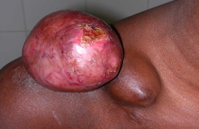
Figure 1: Dermatofibrosarcoma of Darier and Ferrand on the shoulder-Two-lobed aspect.
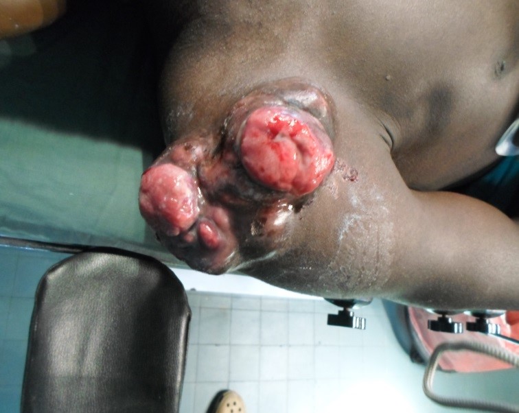
Figure 2: Dermatofibrosarcoma of Darier and Ferrand on the right Shoulder-Ulcerative and budding aspect.
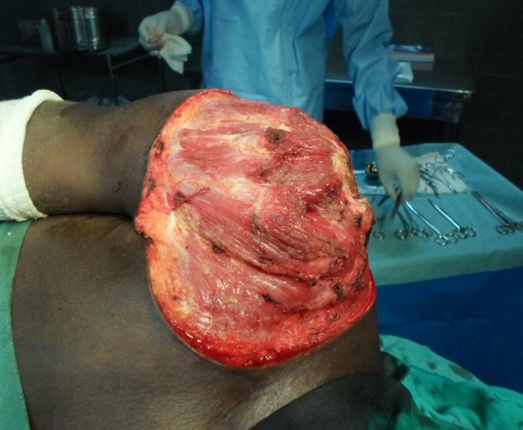
Figure 3: Wide resection with margins of 2,5 cm.

Figure 4:Myocutaneous flap oflatissumusdorsi.
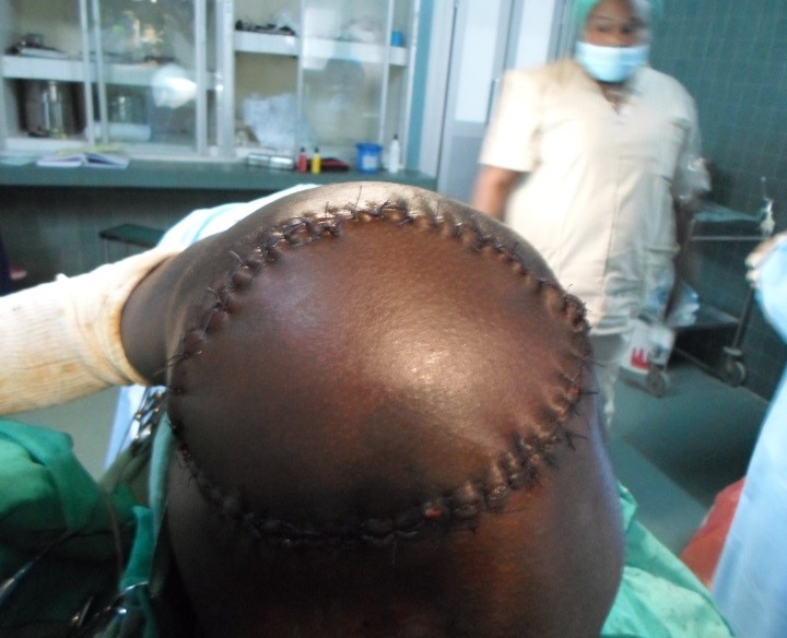
Figure 5: Closure with flap of latissimus dorsi.
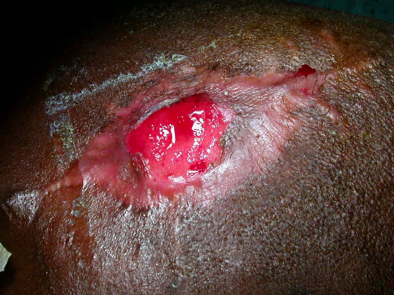
Figure 6: Dermatofibrosarcoma of Darier and Ferrand on the left temporal bone.
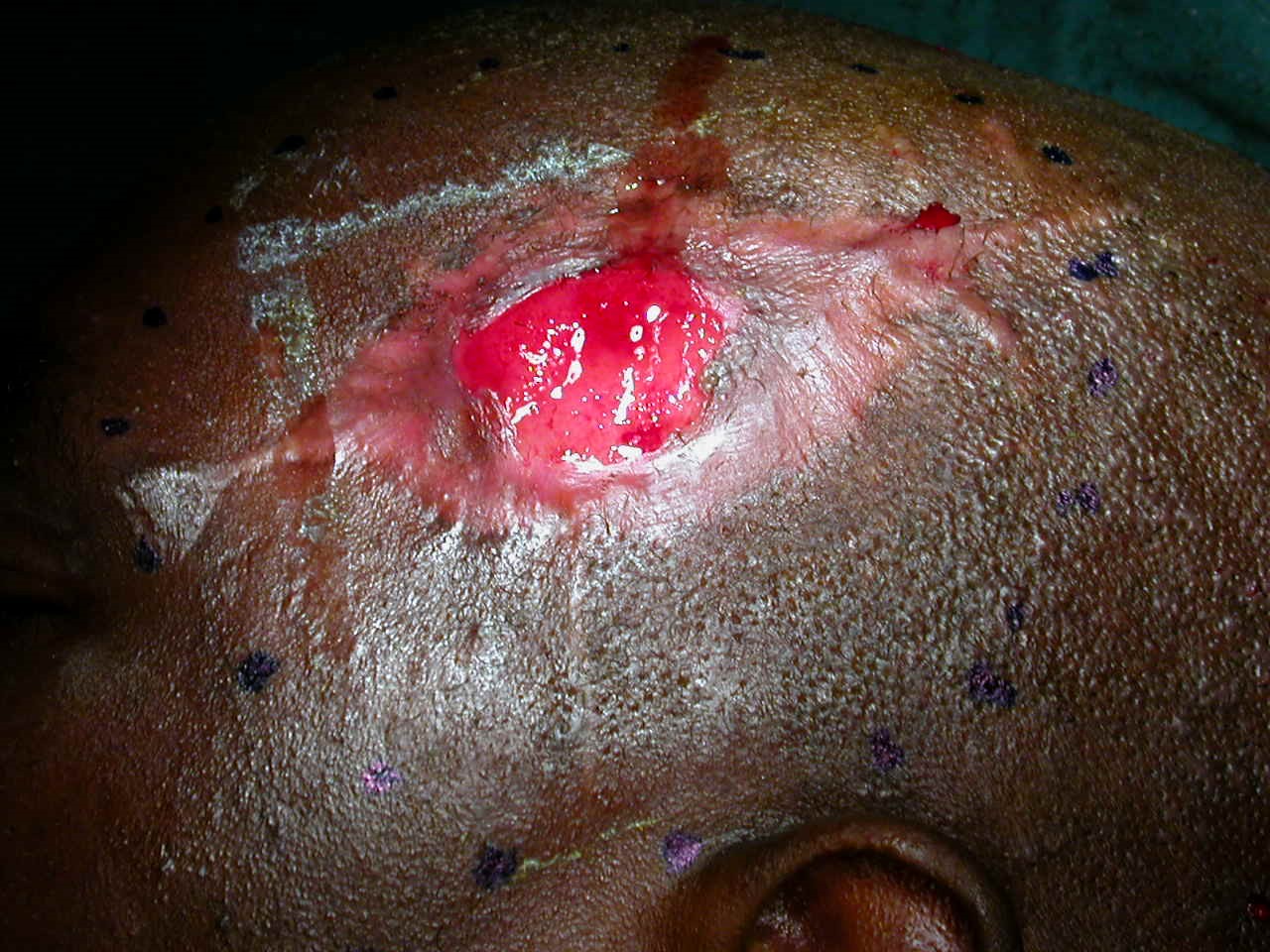
Figure 7: Resection with margins of 2,5 cm
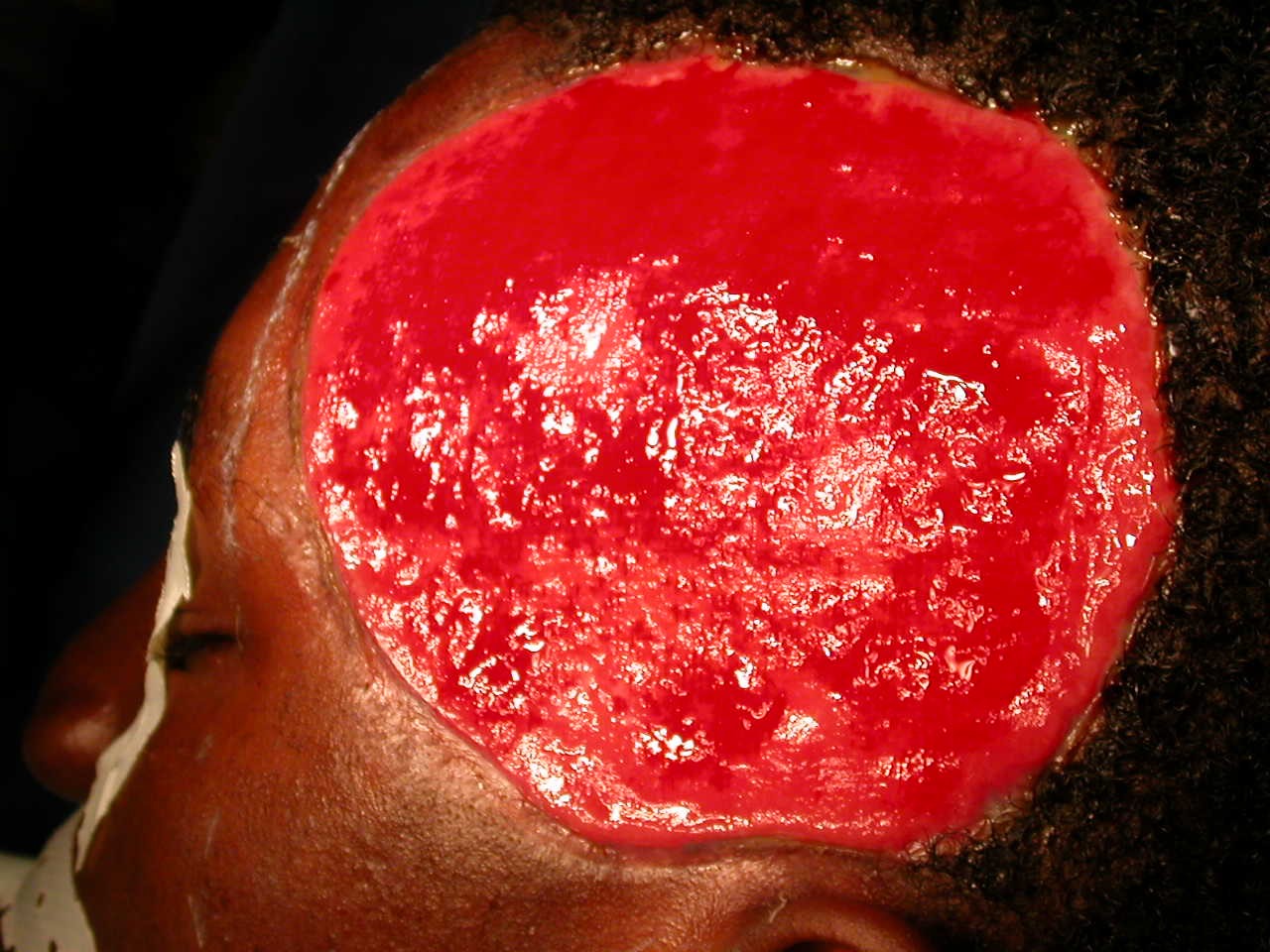
Figure 8:Depth of resection,
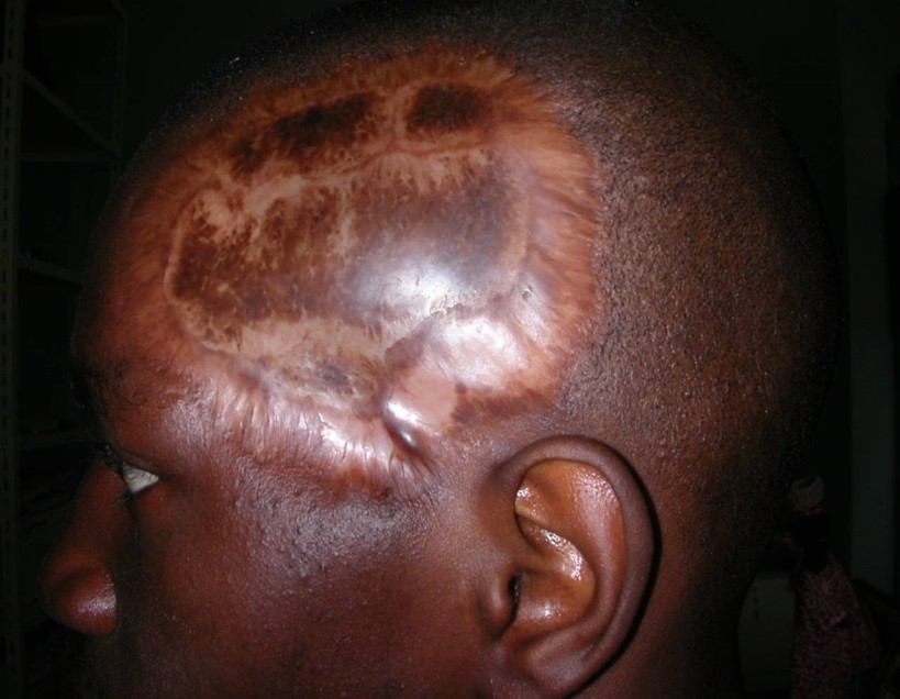
Figure 9: Thin skin graft.
|
Repair modes after surgery |
Number |
% |
|
Direct suture |
2 |
33,3 |
|
Local flap |
2 |
33,3 |
|
Thin skin graft |
2 |
33,3 |
Table 1: Repair modes after surgery.
Citation: Foba L, Sankale Anne-AuroreNdiaye A, Ndiaye L (2020) Darier Ferrand Sarcoma: Clinical and Therapeutic Aspects About 8 Cases. Ann Med &Surg Case Rep: AMSCR-100058
