Journal of Surgery and Insights
Case Report
Gossypiboma, an uncommon cause of Acute Abdomen & Small Bowel Obstruction
Othman Tajoury1,2, Jebril Hwaidi1, Abdulmalik Adel2*
1Aljala hospital, Benghazi, Libya
2The Libyan International Medical University, Benghazi, Libya
*Corresponding author: Abdulmalik Adel, The Libyan International Medical University, Benghazi, Libya, Tel: +00218925953518, E-mail: Makryd324@Gmail.com
Citation: Tajoury O, Hwaidi J, Adel A (2019) Gossypiboma, an uncommon cause of Acute Abdomen & Small Bowel Obstruction. J Surg Insights: JSI-100004
Recieved Date: 06 June, 2019; Accepted Date: 07 June, 2019; Published Date: 11 June, 2019
Abstract
Gossypiboma, textiloma also called Retained Foreign Object (RFO), is one of the surgical complications resulting from missing a surgical gauze inside the abdominal cavity [1]. Unintentional missing foreign body in the abdomen necessitates abdominal re-exploration to retrieve it.This intervention might lead to rises in morbidity and mortality rate of the patient, in addition, long hospital stay and medico-legal issues.
We are reporting a case of a 37-year-old woman who came to our Hospital OPD clinic with abdominal pain. She had a surgical history of open cholecystectomy 18 months back. By clinical examination and radiology investigation, the foreign body was suspected. That is a foreign body further confirmed by abdominal CT. During laparotomy foreign body was found inside the ilium so enterotomy performed and retained surgical gauze retrieved which confirmed the diagnosis of textiloma.
Keywords: Gossypiboma; Retained foreign body; Surgical gauze; Textiloma
Introduction
Gossypiboma, the term is a combined Latin word “Gossypium” means (cotton) and the Swahili “boma” means (hiding place) [2,3]. Illustrates a mass within a patient's body containing a cotton matrix enclosed by a foreign body granuloma [4,5]. "Textiloma" is derived from textile (surgical sponges in the previous decades it was made of fabric), and “oma” - disease, tumor, swelling in Greek. It’s utilized instead of gossypiboma because the synthetic materials recently used instead of cotton [4]. Both are pointed to a foreign body unintentionally left in the patient abdominal cavity during surgery and the secondary reactions developed against it [6]. Surgical gauze left in the body following surgical operation is dangerous but could be avoided [7]. Gossypibomas usually has a similar, clinical and radiological feature of tumors and abscesses collection, with different complications and signs, which make the diagnosis hard to reach [8]. Retained surgical foreign bodies cause two types of complications. Type one is an abscess formation. Type two is ended up with foreign body granuloma [4].Uusually, it's asymptomatic for a long period reaching years in some cases [4].
Case Report
A 37-year-old Female Patient referred to Al-Jalla Hospital with generalized abdominal pain and distention, the pain was started one month ago aggravated by food no special reliving factors, accompanied with nausea and vomiting for 2 days. Not known case of any chronic illness No H/O same illness before systemic review unremarkable, past surgical history: She underwent open Cholecystectomy surgery 18 months ago. General examinations and laboratory investigations were within normal limits. On abdominal examination, Right Kocher incision scar was present. Conservative management was started as for acute abdomen. Plain abdominal X-rayshows a sign of a radio-opaque marker in the lower left abdomen (Figure 1). Abdominal ultrasonography showed dilated small bowel. The abdominal-pelvic computerized tomography (CT) scan, shows intra-abdominal surgical gauze shadow (Figure 2). Exploratory laparotomy was planned under general anesthesia. Through low midline incision, abdominal cavity explored. The mass was found inside the small bowel in the ilium. We took all the precautions to prevent spillage of small bowel contents then we open the ilium in which a sponge (Medium sized abdominal gauze) was found then pulled out (Figure 2&3). The sponge was removed (Figure 4&5) and ilium stitched using Vicryl stitch. The abdomen layers were closed with all safety measures and counts of sponges and instruments. The patient recovery period was well without complains. After 5 days, the patient was discharged and advised to follow-up our OPD clinic.
Discussion
The most significant emergency case is the acuteabdomen that usually needs surgical intervention. Mesenteric Ischemia, Appendicitis, Diverticulitis, Subacute Bowel Obstruction, are some of the most frequent differential diagnosis. An adult patient with an acute abdomen generally looks unwell and has abnormal findings on physical examination. There may be upper and lower gastrointestinal (GI) signs and symptoms associated with signs of Peritonitis. Appropriate management differs from conservative treatment to surgical removal of the definitive cause [5]. Gossypiboma cases generally have subacute or chronic presentations that are different from other acute abdomen cases. Gossypiboma cases may cause the specialist to lose their job. Due to medico-legal inference data regarding the actual occurrence is hard to calculate because of low reporting rate [6]. It differs among 1 out of 1,000-1,500 intra-abdominal operations and 1 out of 300-1,000 of all operation [7]. A Gossypiboma can be difficult to identify just by utilizing radiological screening because in some cases the sponge doesn't have any radiological marker on itself, for example the cotton can simulate a hematoma, granulomatous process, abscess formation, cystic mass or neoplasm in any postoperative patient The possibility of a Retained Foreign Object should be included in the differential diagnosis in case the patient is presented with pain, signs of infection or a palpable mass. Laparoscopic retrieval of retained gauze may be possible in case of early diagnosis [8]. According to Wan et al Gossypibomas were most commonly found in the abdomen (56%), pelvis (18%), and thorax (11%). The main form of detecting a Gossypibomas is a CT scan (61%), other forms of detection include radiography (35%), and ultrasound (34%). Pain or irritation (42%), palpable mass (27%), and fever (12%). (6%) of the cases were found to be asymptomatic. Complications of such cases included adhesions (31%), abscess formation (24%) and fistula formation (20%). Average discovery time was calculated to be 6.9 years with a median (quartiles) of 2.2 years (0.3-8.4 years) [9]. In a case report conducted by Sumer et al. Gossypiboma was extracted from the abdominal cavity 23 years after a cesarean operation, making it the longest time in the literature of foreign bodies. The possible causes of sponge retentions are emergency surgery, difficult surgical procedure, rushed sponge counts, long operations, and inexperienced staff. Most cases were due to false correct counting at the end of surgery. Gossypiboma commonly follows the gynecological and upper abdominal emergency surgical procedures. The diagnosis is usually reached by radiological studies and a high suspicion. The clinical presentations of Gossypiboma can vary depending on the location of the sponge and the type of reaction. The two types of foreign body reactions in pathology are exudate and aseptic fibrinous reaction the exudate reaction can lead to abscess formation, and chronic internal or external fistula formation, while aseptic fibrinous reaction can result in adhesions, encapsulation and granuloma formation. They usually remain asymptomatic or are presented with pseudo-tumour syndrome.
Common signs and symptoms of Gossypiboma include abdominal distention, ileus, tenesmus, pain, a palpable mass, diarrhea, abscess,and fistula formation, with also nausea, vomiting, anorexia and weight loss.
Retained surgical sponges can cause serious complications, like bowel or visceral perforation, obstruction or fistula formation, sepsis or even death. Intraabdominal Gossypibomas can migrate into the ileum, stomach, colon or bladder without any apparent opening in the wall of these luminal organs, like in our case as we found it inside the lumen of ileum instead of the abdominal cavity [9]. The inflammatory granulomatous reaction is the most likely cause of the extra-osseous accumulation of technetium-99m-methylene diphosphonate (Tc-99m MDP) in Gossypibomas as studied by nuclear medicine study. A published article by the New England Journal of Medicine contains the risk factors of Retained Foreign Bodies (RFBs). The authors identified only 3 out of the 8 risk factors including emergency surgery, unexpected change in the operation and body mass index [BMI]. These 3 were found to be statistically important by multivariate logistic regression. The counting of sponges and instruments was not a significant predictor. The most impressive imaging finding of Gossypiboma is the coiled or stripy radio-opaque lines on plain abdominal X-ray. The ultrasound usually shows a well-defined mass containing echogenic focus with a hypoechoic edge and a tough posterior shadow. On CT, a Gossypiboma may manifest as a cystic lesion, hyper-dense capsule, concentric layering, and mottled shadows as bubbles or mottled mural calcifications. MRI characteristics of Gossypiboma in the abdomen and pelvis involve the picture of a well-defined mass with a peripheral wall of low signal intensity on T1- and T2-weighted imaging, with whorled stripes seen in the central portion and peripheral wall enhancement after intravenous gadolinium administration on T1-weighted imaging. Prevention of Gossypiboma can be done by simple precaution, for example, keeping a detailed pack count and labelling the packs with markers. New technologies are being established that might help reduce the incidence of RFP, like radiofrequency chip identification (RFID) by the barcode scanner. The purpose of this system would be to reduce errors in the sponge count by removing the human error factor. Furthermore, the sponge count protocol itself has been involved as a risk to patient safety.
Conclusions
Gossypiboma raised the red flag for every surgeon to be aware of the risk factors that might lead to retained gauze (A gossypiboma) and take precautions to avoid it. Although gossypiboma is rare, it should be well-thought-out in the differential diagnosis of sub-acute intestinal obstruction in patients who underwent laparotomy previously. The best approach in the prevention of this condition can be achieved by a carefulcount of surgical tools in addition to thorough exploration of the surgical site at the end of surgery and also by routine use of surgical sponges which marked with a radio-opaque indicator.
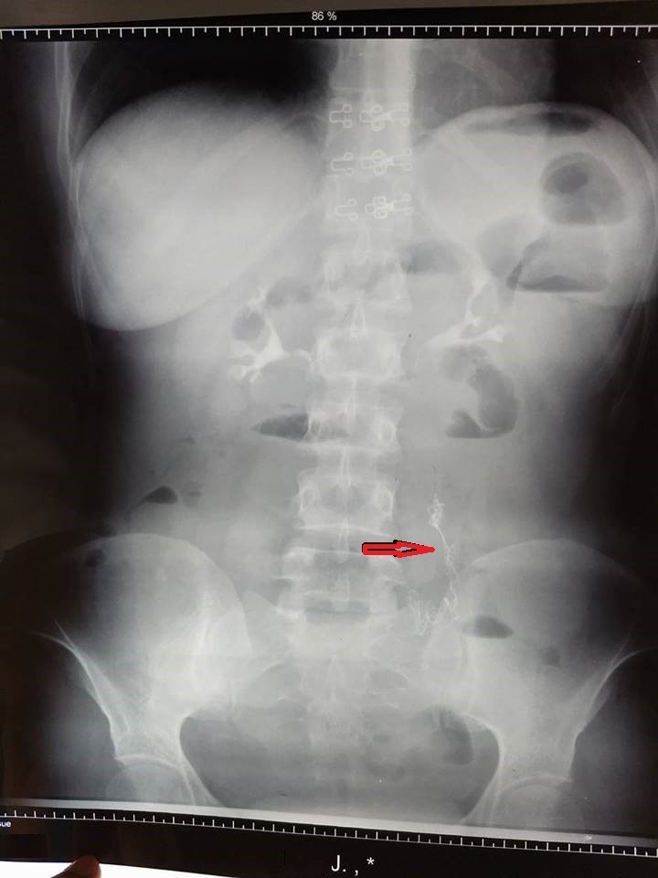
Figure 1: Plain A/P abdominal x-ray showing a sign of a radio-opaque marker in the left lower abdomen.
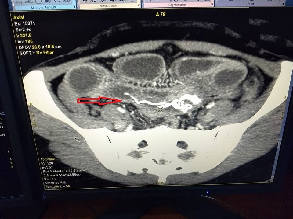
Figure 2: CT scan of the abdominopelvic region showing shows intra abdominal Surgical gauze shadow with a radio-opaque marker.
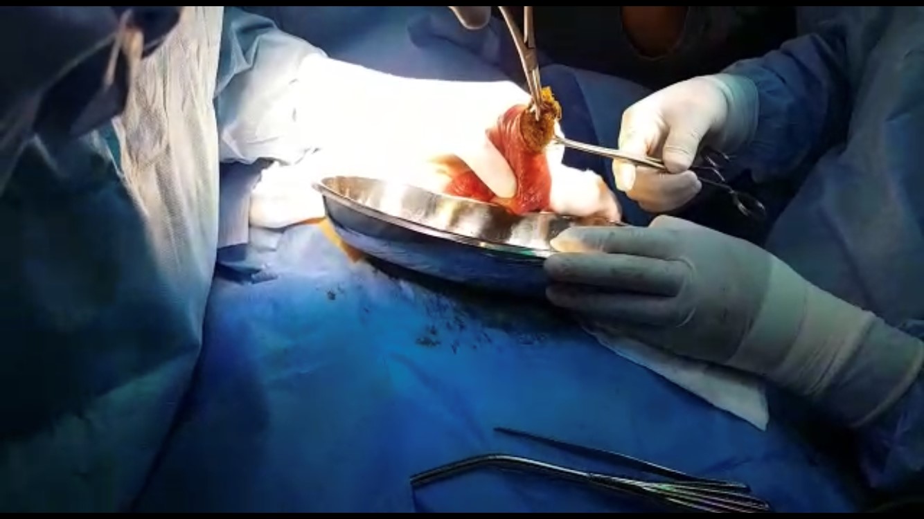
Figure 3: After opening the ilium (holding with Babcock forceps).
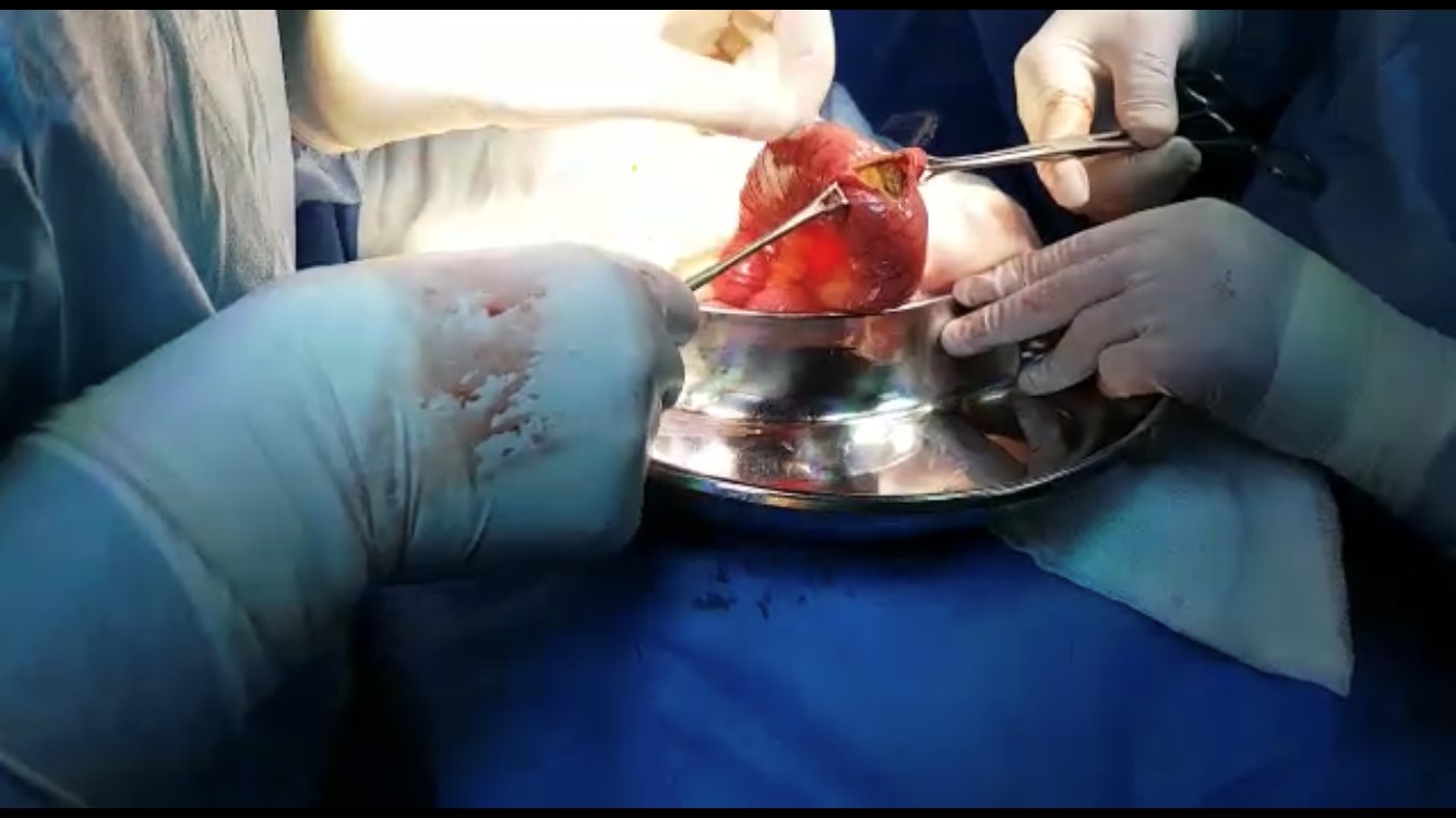
Figure 4: pulling the gauze with Babcock forceps.
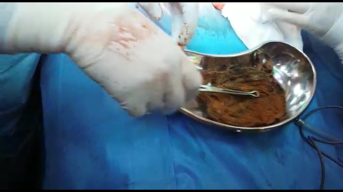
Figure 5: Specimen of a medium-sized abdominal gauze as retained foreign body removed from the small bowel.
Citation: Tajoury O, Hwaidi J, Adel A (2019) Gossypiboma, an uncommon cause of Acute Abdomen & Small Bowel Obstruction. J Surg Insights: JSI-100004.