
Journal of Obstetrics and Gynecological Problems
Case Report
A New Surgical Management in Post-Radiation Gynecological Fistulas
Silva JMM1*, Gonçalves EL2, Melo LHF3, Neto JAV4, Lacerda CA5, Sousa LFPA6, and Brito LA7
1Department of Gynecology and Obstetrics, Universidade de Brasília, Brazil
2Department of Gynecology and Obstetrics, Hospital Geral de Fortaleza, Brazil
3Department of Gynecology and Obstetrics, Universidade de Brasília, Brazil
4Department of Gynecology and Obstetrics, Hospital Geral de Fortaleza, Brazil
5Department of Gynecology and Obstetrics, Hospital da Região Leste, Brazil
6Department of Gynecology and Obstetrics, Fertilcare-Human Reproduction Center, Brazil
7Department of Medicine, Universidade Federal do Ceará, Brazil
*Corresponding author: João Marcos Meneses Silva, Department of Gynecology and Obstetrics, Universidade de Brasília, Brazil, Tel: +556133072028, Fax: +556133403385; Email: drjoaomarcosmeneses@hotmail.com
Citation: Silva JMM, Gonçalves EL, Melo LHF, Neto JAV, Lacerda CA, et al. (2019) A New Surgical Management in Post-Radiation Gynecological Fistulas. J Obstet Gynecol Probl: JOGP 100003.
Received date: 20 August, 2019; Accepted date: 27 August, 2019; Published date: 02 September, 2019
Abstract
The radiotherapy despite its great value in invasive cervical and endometrial cancer causes major complications such as rectum and vesico-vaginal fistulae. Such complications create great challenge for surgeons due to the difficult dissection techniques. In this study we will demonstrate a new surgical technique involving the Martius flap and the gluteal fold. Three patients with rectal or vesico-vaginal fistulae after radiotherapy were submitted to a new procedure, which consisted of this new surgery technique.Two of whom died, one due to strangulated hernia and the other from pulmonary embolism. The remaing one progressed without complications. The new approach consists of a simple Technique, not exposing the patient to extensive surgery. However, we need more cases for comparison.
Keywords: Cervical cancer; Endometrial cancer; Fistulae; Radiotherapy
Abbreviations: BMI: Body mass index
Introduction
Radiotherapy plays an important role in the treatment of invasive carcinoma of the cervix and endometrium, as adjuvant to surgery or as sole therapy. However, damages to surrounding normal tissues (rectum, bladder, small intestine) by irradiation can lead to complications such as enterocolitis, proctitis, Rectal or vaginal perforation and stenosis or even vaginal-vesical fistulae.
The treatment of recto-vaginal fistulae after radiotherapy is a challenge for the surgeon because of the difficult dissection techniques due to poor irrigation and fibrosis of the irradiated tissue, leading to failures in the repair, especially in the larger lesions. We present a new procedure for repair of post-radiation bigger fistulas that combines the transposition of two known flaps; the Martius flap and gluteal crease. Although those transpositions exist in the literature, they haven’t been described to such use [1,2]. The Martius flap is the result of the mobilization of the bulbocavernosus muscle from the large lip, cut in its posterior portion, and supported by lower pedicle, consequently transposed into the vagina through the lateral vaginal wall. The gluteal fold flap of skin measures ten to fifteen centimeters in length by three centimeters wide and nourished by pudendum vessels. It was designed in the shape of ellipse and the gluteal fold as the axis, being the reference points that demarcate the point of rotation, thus transposed to the posterior wall of the vagina by the mid-lateral tunnel [3]. The region of the fistulae is first prepared with debridement of all edges and surrounding mucosa. The fistulous orifice is closed in single plan with suture in bag or separate points. Next step is covered suturing the two flaps-the first of Martius and defatted the flap over the gluteal fold. Next the suture is made of the donor areas.
Materials and Methods
Our series enrolled three patients with cervical cancer who underwent radiotherapy. All patients progressed to the formation of rectovaginal fistulae. The patients were assessed to exclude recurrent malignant disease and radiological investigations were performed to assess the site, extent and severity of damage at the bowel and all of them had a preliminary transverse colostomy. The first patient ESC 79 years’ old who had a BMI of 35 and was treated with radiotherapy alone for squamous cell carcinoma of the cervix in 1994. Four years later the patient was referred to our hospital for treatment of sequelae -rectovaginal fistulae. Two months after colostomy procedure the Martius flap was used associated to gluteal flap fold as a technique for the corrective surgery. After four months, the colostomy was closed and the flap was feasible and there was no sign of any fistulas. It remained viable for six months after the corrective procedure but the follow up was terminated since the patient monitoring was discontinued because the patient died of intestinal perforation after strangled hernia. The second patient NLMS, 54 years, history of radio and chemotherapy, developed recto-vaginal fistulae. The correction using the flap gluteal fold and Martius flap was done with good development of retail. The flap was showing good conditions until two months after surgery and monitoring stopped because the patient died due to pulmonary embolism. The third patient RBP 42 years old, with history of 2 fistulas due to radiotherapy-one rectal-vaginal and other vesico-vaginal. The patient underwent a colostomy procedure and after forty-two days was subjected to the technique of repair with the use of the Martius flap and the gluteal fold in 2007. Until now evolved without complications and with retail clinics with good vitality.
Discussion
Despite the progress in techniques of radiotherapy, there are still urinary and intestinal complications, in some cases serious and challenging to treat. The small fistulas -less than 1 cm may be corrected successfully, only with local vaginal repair with colpocleisis of the upper third part of vagina. However bigger lesions Demand more complex approaches. Some authors have published transabdominal techniques; resecting the irradiated rectum and sigmoid and performed colon-anal anastomosis [3]. Others advocate the interposition of vascularized pedicles between the rectum and vagina, with epiplon or rectus abdominal muscle associated with transabdominal repair of fistulae [4]. Approaches are difficult to implement in an already irradiated pelvis with considerable postoperative morbidity. The technique used is important for its simplicity in implementation, using flaps with good vascular supply and mild scarring sequelae in the donor area (Figure 1-3). In addition, it does not expose the patient to extensive surgery, with high rates of morbidity due irradiation induced changes affecting the pelvis. However, this technique can be criticized for not removing the damaged segment of the rectum by radiotherapy, and cannot be used when there is significant stenosis. Furthermore, more cases should be treated by the method so that results can be validated and compared with other experiments.
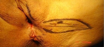
Figure 1: Design of the Martius and the gluteal fold flap before the surgery in a case of rectovaginal fistulae. (Case 1 in our patient serie).
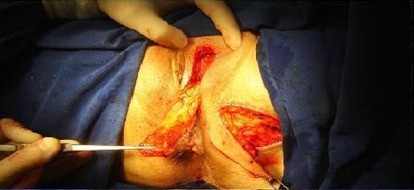
Figure 2: Mobilization of bulbocavernosus muscle from the large lip (Martius flap) and made the gluteal fold flap. (Case 2 in our patient serie).
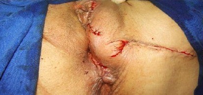
Figure 3: The fistulous orifice and the donor areas were closed in single plan, with suture in separated points. (Case 2 in our patient serie).
Citation: Silva JMM, Gonçalves EL, Melo LHF, Neto JAV, Lacerda CA, et al. (2019) A New Surgical Management in Post-Radiation Gynecological Fistulas. J Obstet Gynecol Probl: JOGP 100003.
