
Insights of Cardiology Open Access
Case Report
Absent Pulmonary Valve Syndrome with Common Atrioventricular (AV) Valve: A Rare Association
Swati Kapoor1*, Neeraj Awasthy2, Rajeev Upreti3
1Department of Internal Medicine, Max Super Speciality Hospital, New Delhi, India
2Department of Pediatric Cardiology, Max Super Speciality Hospital, New Delhi, India
3Registrar in George Eliot hospital,United kingdom, UK
*Coressponiding author: Swati Kapoor, Department of Internal Medicine, Max Super Speciality Hospital, 1 Press Enclave Road, Saket, New Delhi, India, Tel: +918800861884, E-mail: swatikapoor3012@gmail.com
Citation: Kapoor S, Awasthy N, Upreti R (2019) Absent Pulmonary Valve Syndrome with Common Atrioventricular (AV) Valve: A Rare Association. Insights Cardiol Open Acc: ICOA-100003
Received date: 30 April, 2019; Accepted date: 17 June, 2019; Published date: 25 June, 2019
Abstract
Absent pulmonary valve syndrome (APVS) is a rare congenital anomaly characterized by dysplasia or absent leaflet tissue associated with significant incompetence of the pulmonary valve; pulmonary annular stenosis. It is associated with many congenital anomalies. Here in we report its rare association with common Atrioventricular (AV) valve.
Keywords: Absent pulmonary valve; Common Atrioventricular valve; Intrauterine
Introduction
Absent pulmonary valve syndrome (APVS) is characterized by dysplastic or absent pulmonary valve, free pulmonary regurgitation, and annular stenosis [1,2]. APVS accounts for 0.2-0.4% of live born infants with congenital heart disease [3] and is associated with many congenital anomalies. However we report a rare association of APVS with common atrioventricular (AV) valve which has not yet been reported in literature.
Case Report
A 31 year old multigravidarum presented to pediatric cardiology department at 32 weeks and 6 days of pregnancy. She was referred by her obstetrician on suspicion of fetal heart anomaly on ultrasound. Fetal echocardiography revealed situs solitus, levocardia, and normal pulmonary and systemic venous drainage. There was common Atrioventricular (AV) valve ( Figure 1), double outlet of right ventricle with more than 60% aortic override, sub aortic ventricular septal defect (VSD) and normally related systemic vessels there was an absent pulmonary valve with hypoplastic pulmonary annulus (PA). Pulmonary stenosis had a gradient of 32mm Hg (PA -4.2mm, aortic annulus-7.8mm) with free pulmonary regurgitation. There were centrally dilated pulmonary arteries (right pulmonary artery 8mm and left pulmonary artery 7.8mm) ( Figure 2 and 3) . This turned out to be a rare combination of APVSwith common AV valve.
Discussion
APVS is a rare congenital anomaly characterized by dysplasia or absent leaflet tissue associated with significant incompetence of the pulmonary valve, pulmonary annular stenosis. This often leads to rise in pulmonary artery pressures causing varying degree of airway compression at the tracheal and major bronchial arborization [4,5].
Prognosis depends on the degree of airway compression, a feature that is difficult to diagnose before delivery.
In study by D.Wertaschnigg et al, 12 cases diagnosed between 2000 and 2010 were studied. Ten of the cases(83%) had Fallot type APVS. The other two had isolated APVS and tricuspid atresia respectively [6]. Another case series of 36 APVS patients by Martin A. Nørgaard et al had 97% of them to be variant of Fallot type. Eleven patients (31%) with this morphology had associated vascular abnormalities. Two patients (5%) had DiGeorge syndrome.While one had APVS with double outlet right ventricle (DORV), double committed ventricular septal defect (VSD), and the aorta anterior to the main pulmonary artery [7].
After detailed research of literature, we could not find any case with combination of APVS and common AV valve as in our case. Thus making it a unique association in APVS patient.We presume APVS diagnosed intrauterine has poor prognosis and the combination with DORV is less likely to survive in view of severe compromised right ventricle which effectively serves as a systemic ventricle in the foetus.
Also as per study conducted by Gottschalk et al APVS diagnosed in the first trimester is significantly associated with Tetralogy of Fallot(TOF), patency of the ductus arteriosus(PDA), abnormal Doppler parameters, lethal trisomies and intrauterine mortality. Cases of APVS with isolated TOF and agenesis of the DA have a better outcome than those with additional anomalies, with > 80% survival. Thus further dwelling on importance of regular obstetrician follow ups as well as on diagnostic importance of fetal echocardiography [8]. PDA was absent in our case.
Newborns with isolated APVS may also develop congestive heart failure after birth due to abundant left-to-right shunting over a persistently large ductus arteriosus. Urgent duct ligation will alleviate the hemodynamic situation [9]. The course of isolated APVS during childhood is reportedly benign, with most patients remaining asymptomatic [10].
Hence by this case we would like to report a rare association of common AV valve with APV which is not yet cited in literature
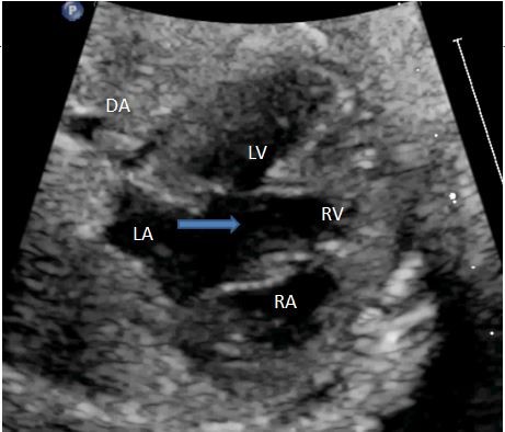
Figure 1: Two-dimensional echocardiography with apical 4 chamber view showing common AV valve (marked by arrow) with unbalanced AVSD (atrioventricular septal defect).
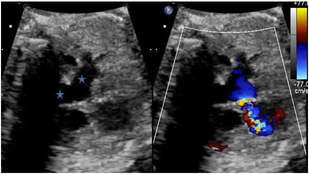
Figure 2: Two- dimensional echocardiography with color compare showing dilated pulmonary artery branches with annular hypoplasia.
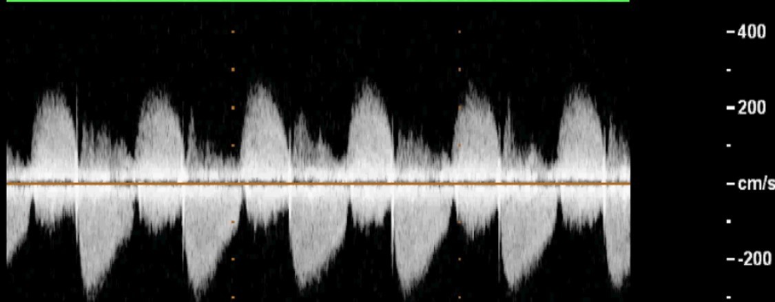
Figure 3: Doppler velocity across RVOT showing free PR with flow acceleration across RVOT with peak velocity of 300/s.
Citation: Kapoor S, Awasthy N, Upreti R (2019) Absent Pulmonary Valve Syndrome with Common Atrioventricular (AV) Valve: A Rare Association. Insights Cardiol Open Acc: ICOA-100003.
