Emergency Medicine and Trauma Care Journal
Case Report
Lingual and Sublingual Haematoma Post Gastroscopy: A Case Report
Rahmani SH1, Dehkharghani MZ 2 and Pouryahya P3,4*
1Department of Emergency, Ostad Alinasab Hospital, Iranian Social Security, Tabriz, Iran
2Department of Emergency Medicine, Shams Hospital, Tabriz University of Medical Sciences, Tabriz, Iran
3Department of Emergency, Casey hospital, Monash Health, Victoria, Australia
4Department of Medicine, Nursing and Health Sciences, Monash University, Victoria, Australia
*Corresponding author: Pourya Pouryahya, Department of Medicine, Nursing and Health Sciences, Monash University, Victoria, Australia, Tel: +61 (03) 8768 1869; E-mail: Pourya.Pouryahya@monashhealth.org
Citation: Rahmani SH, Dehkharghani MZ, Pouryahya P (2019) Lingual and Sublingual Haematoma Post Gastroscopy: A Case Report. Emerg Med Truama. EMTCJ-100017
Received date: 05 November, 2019; Accepted date: 12 November, 2019; Published date: 19 November, 2019
Abstract
Lingual and sublingual haematoma without any associated trauma or any bleeding risk factors are rare entities and commonly described in patients on anticoagulation therapy. An 81-year-old female presented to our emergency department with a 2-hour- history of tongue swelling. Her medical history consisted of non-medicated hypertension without any coagulopathy. She was not taking any medication and had not underwent a dental work up. However, she underwent a semi urgent gastroscopy 2 weeks prior. The examination of the oral cavity revealed a mildly tender haematoma on dorsum and ventral surface of the tongue as well as the floor of her mouth. Her blood test parameters were within normal limits. She was admitted to otolaryngology ward under close observation for further investigations and probably potentially life-threatening airway obstruction. She remained stable without any progression of her swelling and was discharged home after 2 days without any intervention. On review 1 week later, there was a significant reduction in her haematoma size. In spite of few case reports of such haematoma in hypertensive patients, we conclude that in this case delayed lingual and sublingual haematoma following upper GI endoscopy should be considered as her blood pressure was not severely elevated.
Keywords: Complications; Gastroscopy; Haematoma; Lingual; Tongue
Introduction
Case presentation
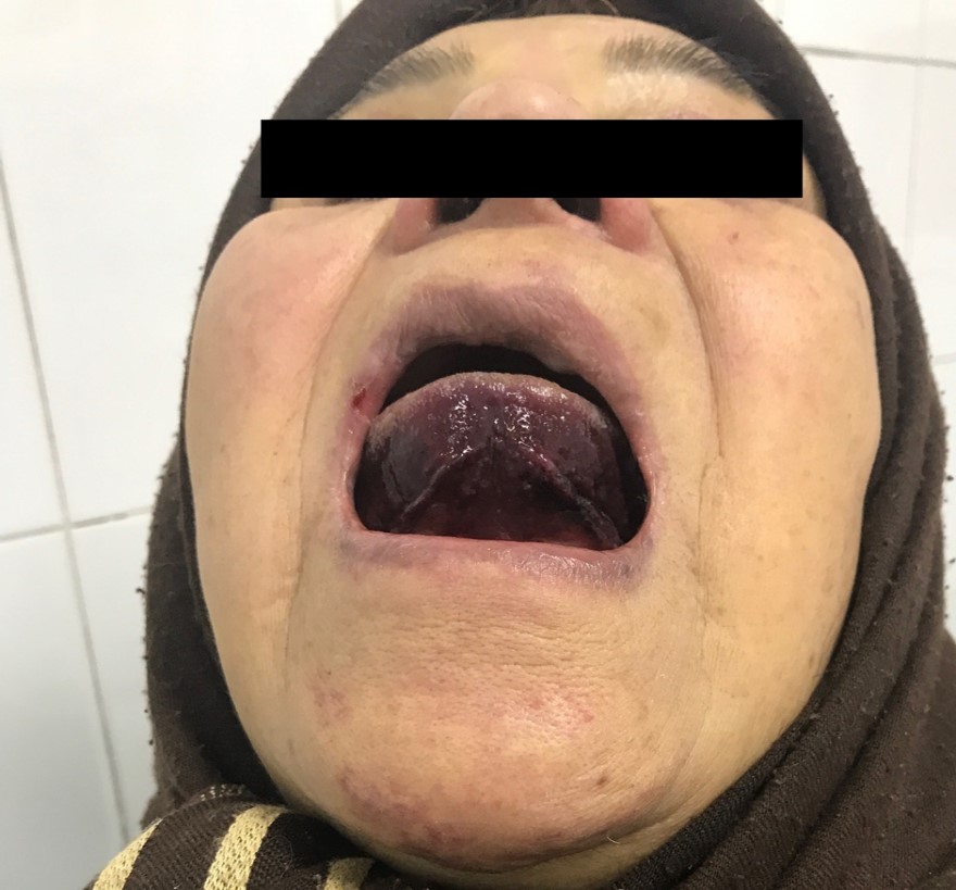
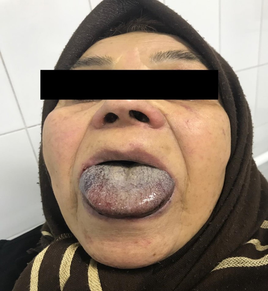
An 81-year-old female presented to the emergency department with a 2-hour history of tongue swelling. Her medical history consisted of non-medicated hypertension without any coagulopathy. She was not taking any medication and had not underwent a dental work up. However, she was admitted for Upper GIB secondary to peptic ulcer disease requiring blood transfusion (2 PRBC) and semi urgent gastroscopy 2 weeks prior. Apart from discomfort in her mouth and inability to completely close her mouth secondary to swollen tongue, she was otherwise asymptomatic. Her airway was patent, and she had no complaint of any respiratory symptoms. The patient was apyrexial, alert and oriented. Her blood pressure was 160/90 mm Hg, heart rate was 100 beats per minute with normal rhythm, respiratory rate was 21 breaths per minute and O2 sat was 96% (in room air). The examination of the oral cavity revealed a mildly tender haematoma on dorsal and ventral surface of the tongue as well as floor of the mouth (Figure 1,2). The swelling was slightly visible over submental area and the anterior neck. Her blood test parameters including platelet and coagulation profile were within normal limits. She was admitted to otolaryngology ward for close observation and further investigations. She remained stable without any progression of her swelling and was discharged home after 2 days without any complications or interventions. On one-week follow-up post discharge, she remained well, with significant reduction in her haematoma size.
Discussion
Although spontaneous lingual and sublingual haematoma are rare entities but always should be consider as a potentially airway and life-threatening conditions. They can occur in elderly patients and the theory behind is aneurismal changes in the facial and lingual arteries. It seems hypertensive patients probably are susceptible to rupture of these weak vessels and subsequent haematoma [1-4]. The lingual artery and its branches supply tongue well and any bleeding from these arteries can cause a severe haematoma [5]. Furthermore, as the floor of the mouth contained of several potential spaces which are covered by some loose tissue, any bleeding can cause an expanding haematoma and airway obstruction6. However, such haematoma following dental procedures or anticoagulation therapy are not rare [2-4] reported, a 45-year-old female who underwent placement of four implants in her anterior mandible. During the procedure, she demonstrated a severe pain, signs of airway obstruction, and dysarthria. Computerized tomography revealed a massive lingual and sublingual haematoma. After 2 days of conservative management, the signs and symptoms had resolved completely. They concluded that demonstrating the mandibular atrophy, carefully selection of the correct length and angulation of bone drilling and keeping attention to the flapless technique should be exactly considered in high risk cases [3] reported, a 70-year-old woman with a 2-day-history of sublingual and lingual haematoma who was taking warfarin over the last 2 weeks. Upon arrival, she was stable without any difficulty in breathing. On examination, there was a soft and red haematoma in the ventral surface of tongue as well as the floor of the mouth. INR was markedly elevated; therefore, fresh frozen plasma and vitamin K commenced. Three days later, the haematoma had disappeared entirely. That was the first isolated lingual and sublingual haematoma following a short time of using warfarin [5] reported, a case of progressive sudden-onset sublingual haematoma in a hypertensive patient, who had no history of trauma, recent dental work or taking anticoagulants. On arrival, the patient was tachypneic with a blood pressure of 220/100. Laboratory tests were within normal limits and CT angiography was normal [6] and Shouvanik Satpathy et all in 2015 reported, two similar cases of submental haematoma in hypertensive patients without any history of recent trauma or taking anticoagulants. CT scan showed no AVM or pseudoaneurysm of lingual artery. In all 3 case reports the authors concluded the rupture of supplying atherosclerotic vessels due to uncontrolled sever hypertension as the underlying causes [7-8].
It seems in spontaneous lingual and sublingual haematoma cases, the only required intervention is electively securing the airway and controlling blood pressure. The team should be experienced in surgical airway management and performing fiberoptic technique. Our case was managed conservatively without any emergent intervention in her blood pressure as the blood pressure was not severely elevated and decreased slightly after admission and reassurance.
Conclusion
Although there are few case reports of lingual haematoma in patients with uncontrolled hypertension, but most iatrogenic cases occur after dental implants placement and thrombolytic therapy. However, we conclude that in this case delayed lingual and sublingual hematoma following upper GI endoscopy should be considered as her blood pressure was not severely elevated.
Acknowledgement
Special thanks to the emergency and otolaryngology Departments staff of the Ostad Alinasab Hospital of Tabriz.

Figure 1 : Haematoma on ventral surface of the tongue.

Figure 2 : Haematoma on dorsal surface of the tongue.
Citation: Rahmani SH, Dehkharghani MZ, Pouryahya P (2019) Lingual and Sublingual Haematoma Post Gastroscopy: A Case Report. Emerg Med Truama. EMTCJ-100017.